Home

elevated umbilical artery dopplers

Intrauterine growth restriction - abnormal umbilical artery Doppler |  Radiology Case | Radiopaedia.org
Intrauterine growth restriction - abnormal umbilical artery Doppler | Radiology Case | Radiopaedia.org

Doppler assessment of the fetus with intrauterine growth restriction -  ScienceDirect
Doppler assessment of the fetus with intrauterine growth restriction - ScienceDirect

Figure 5 from Examination of the fetal cardiovascular system. | Semantic  Scholar
Figure 5 from Examination of the fetal cardiovascular system. | Semantic Scholar

Umbilical Artery Doppler Ultrasound Normal Vs Abnormal Image Appearances |  Spectral Doppler USG - YouTube
Umbilical Artery Doppler Ultrasound Normal Vs Abnormal Image Appearances | Spectral Doppler USG - YouTube

Doppler ultrasound data of the study individuals. Doppler recordings... |  Download Scientific Diagram
Doppler ultrasound data of the study individuals. Doppler recordings... | Download Scientific Diagram
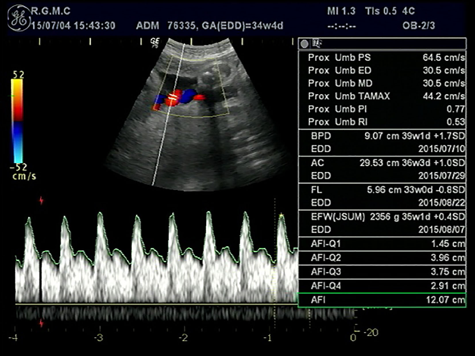
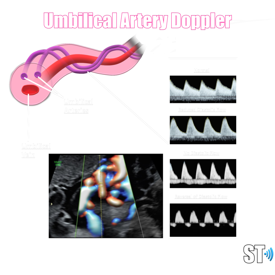
Umbilical Artery Doppler – Sonographic Tendencies

Umbilical Artery Doppler Assessment: A How-To Guide | Empowered Women's  Health
Umbilical Artery Doppler Assessment: A How-To Guide | Empowered Women's Health

Doppler Ultrasonography in Obstetrical Practice - ppt download

Umbilical Artery Doppler – Sonographic Tendencies

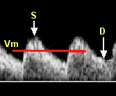
Umbilical Artery Doppler Reference Ranges

32 weeks, 3 days: On becoming an inmate | lamenting the lentil

A Radiologist's Guide to the Performance and Interpretation of Obstetric  Doppler US | RadioGraphics
A Radiologist's Guide to the Performance and Interpretation of Obstetric Doppler US | RadioGraphics

Clinical Applications for Doppler Ultrasonography in Obstetrics | Current  Radiology Reports
Clinical Applications for Doppler Ultrasonography in Obstetrics | Current Radiology Reports

Notching in the Umbilical Artery Doppler Waveform in the Absence of Cord  and Placental Structural Abnormalities: A Case of Massive Fetomaternal  Hemorrhage | Nishimura | Journal of Clinical Gynecology and Obstetrics
Notching in the Umbilical Artery Doppler Waveform in the Absence of Cord and Placental Structural Abnormalities: A Case of Massive Fetomaternal Hemorrhage | Nishimura | Journal of Clinical Gynecology and Obstetrics

PDF] Reference ranges for serial measurements of umbilical artery Doppler  indices in the second half of pregnancy. | Semantic Scholar
PDF] Reference ranges for serial measurements of umbilical artery Doppler indices in the second half of pregnancy. | Semantic Scholar

The clinical application of Doppler ultrasound in obstetrics - Mone - 2015  - The Obstetrician & Gynaecologist - Wiley Online Library
The clinical application of Doppler ultrasound in obstetrics - Mone - 2015 - The Obstetrician & Gynaecologist - Wiley Online Library
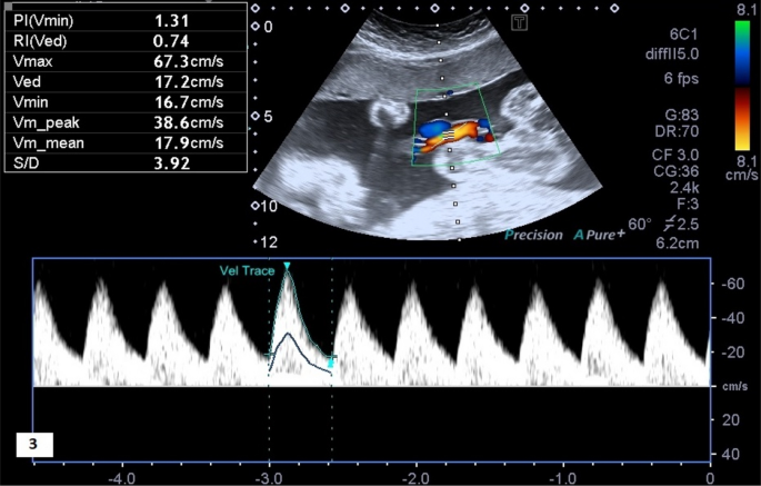
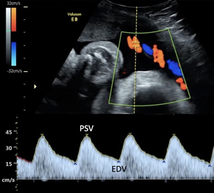
Normal umbilical artery doppler values in 18–22 week old fetuses with  single umbilical artery | Scientific Reports
Normal umbilical artery doppler values in 18–22 week old fetuses with single umbilical artery | Scientific Reports

Fetal ultrasound technique does not improve prediction of  small-for-gestational age babies - USF Health NewsUSF Health News
Fetal ultrasound technique does not improve prediction of small-for-gestational age babies - USF Health NewsUSF Health News

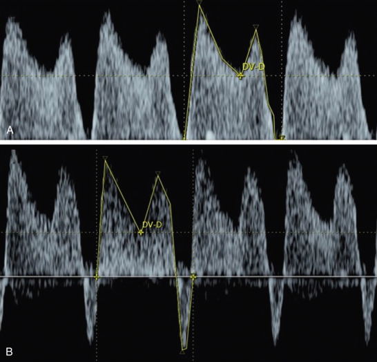
Doppler Ultrasound Evaluation of the Fetus and Placenta | Radiology Key

Reflected hemodynamic waves influence the pattern of Doppler ultrasound  waveforms along the umbilical arteries. - Abstract - Europe PMC
Reflected hemodynamic waves influence the pattern of Doppler ultrasound waveforms along the umbilical arteries. - Abstract - Europe PMC

Normal umbilical artery doppler values in 18–22 week old fetuses with  single umbilical artery | Scientific Reports
Normal umbilical artery doppler values in 18–22 week old fetuses with single umbilical artery | Scientific Reports

Evaluation and Management of Suspected Fetal Growth Restriction -  Obstetrics and Gynecology Clinics
Evaluation and Management of Suspected Fetal Growth Restriction - Obstetrics and Gynecology Clinics

Doppler velocimetry discordance between paired umbilical artery vessels and  clinical implications in fetal growth restriction - American Journal of  Obstetrics & Gynecology
Doppler velocimetry discordance between paired umbilical artery vessels and clinical implications in fetal growth restriction - American Journal of Obstetrics & Gynecology

Abnormal pulsed and color Doppler of umbilical artery with reversed... |  Download Scientific Diagram
Abnormal pulsed and color Doppler of umbilical artery with reversed... | Download Scientific Diagram

Starship - Antenatal Doppler Studies in High Risk Pregnancies

Abnormal Fetal Umbilical Cord Doppler? What Should I Do Next? A Case Study  Demonstrating Corroboration of Umbilical Cord Doppler and Middle Cerebral  Doppler - Michelle M. Morrissette, 2012
Abnormal Fetal Umbilical Cord Doppler? What Should I Do Next? A Case Study Demonstrating Corroboration of Umbilical Cord Doppler and Middle Cerebral Doppler - Michelle M. Morrissette, 2012

Doppler Ultrasound Evaluation of the Fetus and Placenta | Radiology Key